Cell Health Questions Never Stop, Neither Should The Answers.
Whether you’re running a quick viability screen or building a complete picture of cell health, the next step depends on the question you’re asking. Explore our full cell health portfolio and find the right assay for your specific question.
Cell Viability
Are My Cells Alive?
Viability, the number of living cells in a sample, is a question researchers return to at every stage, from primary screen through lead optimization. Viability screens are the normalizing control for proliferation, apoptosis, and pathway-level readouts. An assay that lacks sensitivity at low cell numbers or introduces variability between plates, undermining every downstream result.
Common pitfalls with cell viability assays are:
- Assays lack sensitivity threshold required for reproducibility and high-throughput screening
- Lacking an option for kinetic monitoring
- Viability assays measure how many cells are alive, but not whether they’re actively dividing; additional methods are often needed
Questions & Solutions
How can I measure viability?
The gold standard for measuring viability is the CellTiter-Glo® Viability Assay:
- Single-reagent, single-step ATP detection
- Most-cited viability assay in peer-reviewed research
- Enables scaling for high-throughput screening
Can I track viability in real time?
Monitor viability continuously with the RealTime-Glo™ MT Cell Viability Assay:
- Non-lytic — read the same well over hours or days
- Compatible with downstream multiplexing
How can I decrease my prep time?
Try the CellTiter-Glo® 2.0 Cell Viability Assay for shorter prep time:
- Ready-to-use
- Same trusted CTG chemistry
How can I measure viability but keep my cells intact?
The CellTiter-Fluor™ Assay and CellTiter-Blue® Assay measure viability without lysing cells.
- CellTiter-Fluor™ Assay: Cell-permeable fluorescent, multiplex in the same well
- CellTiter-Blue® Assay: Resazurin-based, works on standard fluorescence readers
Looking for more cell viability assays?
Cell Proliferation
Are My Cells Proliferating?
Cell proliferation, when cells are alive and capable of replication, is a common measurement for researchers trying to understand the health of their cells. A compound that keeps cells alive but stops them from replicating looks like a pass in a viability screen, but that’s a missed signal you need to catch. Proliferation measurements close that gap, separating true cell growth from survival and giving you a clearer read on compound mechanism.
Common limitations with cell proliferation assays are:
- Methods take hours to days to complete
- Assays cannot be reliably scaled up for high-throughput screening
- Measurements require specialized equipment
Questions & Solutions
Are my cells dividing?
Add-Mix-Measure proliferation with the Lumit® Cell Proliferation (Human Ki-67) Assay:
- Increase confidence - A large assay window provides clear separation between treated and control samples
- Reduce lab time and effort using an easy, no-wash, add-and-read protocol
- Faster answers - hKi-67 levels show significant changes at earlier time points, shortening the time to decision
- Clearly discern between changes in proliferation and effects due to cytotoxicity
Is my treatment affecting cell growth?
Determine number of viable cells in proliferation, cytotoxicity or chemosensitive assays with CellTiter 96® AQueous One Solution Cell Proliferation Assay (MTS):
- Fast reliable results with any absorbance reader
- Compare results to decades of published literature
Looking for more cell proliferation assays?
|
Cell Viability (ATP) |
3D Cell Viability (ATP) |
Real-Time Viability |
Non-Lytic Viability |
Cell Proliferation |
|
|---|---|---|---|---|---|---|
| Best Use | Routine viability & HTS screening | Established protocols using original formulation | 3D spheroids, organoids, microtissues | Kinetic monitoring over time; downstream multiplexing | Multiplex first step; cells needed for follow-up assays | True proliferation readout independent of metabolism |
| Key Decision Points | ||||||
| Measures | Viability | Viability | Viability (3D) | Viability (kinetic) | Viability | Proliferation |
| Cells alive after assay? | âś— Lytic | âś— Lytic | âś— Lytic | âś“ Non-lytic | âś“ Non-lytic | âś— Lytic |
| Multiplexing compatible? | LimitedLytic—must be terminal step | LimitedLytic—must be terminal step | LimitedLytic—must be terminal step | âś“ ExcellentNon-lytic; pair with any downstream assay | âś“ ExcellentNon-lytic; pair with Caspase-Glo®, CTG, etc. | ModerateCan multiplex with CellTox™ Green or other fluorescent readouts |
| Real-time monitoring? | âś— Endpoint | âś— Endpoint | âś— Endpoint | âś“ Up to 72hRead same wells repeatedly | âś— Endpoint | âś— Endpoint |
| 3D culture compatible? | PartialWorks for small spheroids; use 3D version for dense structures | PartialSame as 2.0 | âś“ OptimizedEnhanced lysis for dense 3D structures | PartialSubstrate must penetrate; best for small/loose 3D models | PartialSubstrate access may be limited in dense 3D | âś“ YesDetects Ki-67 in cell lysates from any culture format |
| Assay Attributes | ||||||
| Assay Principle | ATP quantitation (luciferase/luciferin) | ATP quantitation (luciferase/luciferin) | ATP quantitation (enhanced lysis for 3D) | Metabolic reduction of pro-substrate to luciferase substrate | Live-cell protease activity (GF-AFC cleavage) | Ki-67 immunodetection via NanoBiT® complementation |
| Detection Mode | Luminescence | Luminescence | Luminescence | Luminescence | Fluorescence400Ex / 505Em | Luminescence |
| Reagent Format | Ready-to-use liquid | Buffer + lyophilized substrateRequires reconstitution | Ready-to-use liquid | 2 components(enzyme + substrate) | Single reagent | Antibody mix + detection reagent |
| Time to Result | 10min | 10min | ~30min | ContinuousFirst read: 1–2h after addition | 30min | ~2h |
| Practical Considerations | ||||||
| Plate Formats | 96, 384, 1536 | 96, 384, 1536 | 96, 384 | 96, 384 | 96, 384 | 96, 384 |
| HTS Suitability | âś“ Excellent1536-well capable; fast protocol | âś“ Excellent1536-well capable | âś“ Good | ModerateRequires kinetic reader scheduling | âś“ Good | âś“ Good |
| Sensitivity (96-well) | ~15 cells/well | ~10 cells/well | Spheroid-dependent | <100 cells/well | ~40 cells/well | Cell line-dependent |
Cell Death and Stress
Cell death can result from different pathways, and oxidative stress can be an upstream trigger. Understanding which process is involved requires different measurements.
How, When, and Why Are My Cells Dying?
When a treatment kills cells, the next question is always how and why. The mechanism determines whether a hit advances or gets dropped, whether toxicity is on-target or off, and what downstream immune and inflammatory responses to expect. No single method tells the whole story: each marker captures one slice of a complex, multipathway process (e.g., caspase activity, membrane changes, DNA fragmentation, or phosphatidylserine exposure). Multiple methods are often needed to confidently identify the mechanism of death.
Common pitfalls with cell viability assays are:
- Traditional workflows are labor-intensive and difficult to scale
- Wash steps, sample manipulation and specialized instrumentation limit throughput and add variability
- Single endpoint measurements can miss the activation window and misrepresent what’s actually happening
Questions & Solutions
Is my compound toxic to cells?
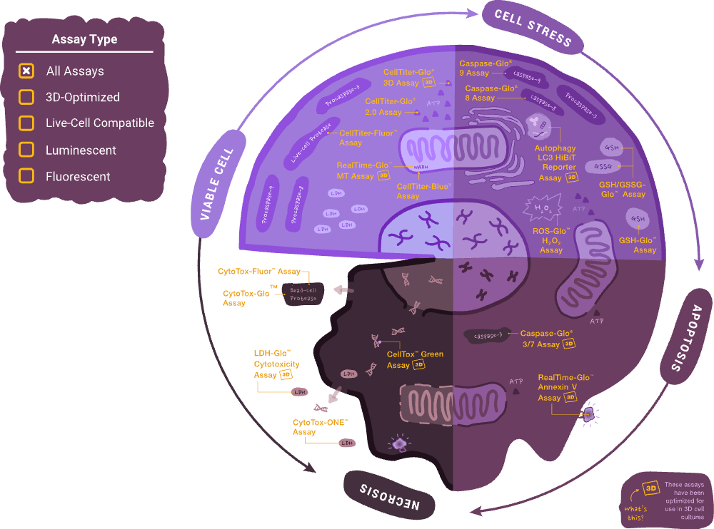
CellTox™ Green Assay detects membrane integrity changes in real time. Signal increases only when membranes are compromised. Multiplex-compatible with CellTiter-Glo® Assay and Caspase-Glo® Assay.
Is my compound triggering apoptosis?
Caspase-Glo® Assays identify which pathways are active. RealTime-Glo™ Annexin V Apoptosis and Necrosis Assay track early apoptosis in real time.
- Caspase-Glo® 3/7 Assay: Executioner caspase activity
- Caspase-Glo® 8 and Caspase-Glo® 9 Assays: Extrinsic vs. intrinsic pathway
- Annexin V Assay: Kinetic monitoring without lysis
Can I measure viability, cytotoxicity and apoptosis in the same well?
ApoTox-Glo™ Triplex and Mitochondrial ToxGlo™ Assays combine readouts in one assay. Triplex: Three answers from one well. Mitochondrial ToxGlo™ Assay: Membrane integrity + ATP viability.
When is cell death occurring?
RealTime-Glo™ Annexin V Apoptosis and Necrosis Assay for kinetic detection of early apoptosis and secondary necrosis in the same well.
RealTime-Glo™ Extracellular ATP Assay monitors ATP release as a real-time indicator of cell membrane breakdown.
With the CellTox™ Green Assay, fluorescent signal increases as membranes are compromised, compatible with kinetic reads over hours or days
LDH-Glo™ Cytotoxicity Assay allows luminescent LDH detection for membrane integrity
Looking for more cell apoptosis assays?
Are my cells under oxidative stress?
Oxidative stress, an imbalance between the production of reactive oxygen species (ROS) and a cell’s antioxidant defenses, is associated with human diseases as well as aging.
Common pitfalls with traditional methods are:
- Probes lack specificity, reacting with multiple reactive species and generating ambiguous results
- Measurements require specialized instruments or complex sample preparation
- Results are difficult to quantify and reproduce between experiments
Questions & Solutions
Is glutathione redox balance shifting?
GSH/GSSG-Glo™ Assay quantifies total glutathione and glutathione ratios in a homogeneous format, giving you a read on whether your cells’ antioxidant defenses are compromised.
GSH-Glo™ Glutathione Assay measures glutathione levels using a homogeneous, bioluminescent readout.
Is nitrite signaling involved?
Griess Reagent System measures nitrite, a physiological messenger in many biological systems.
What stress signaling pathways are activated?
Stress Signaling Pathway Analysis Firefly Luciferase Vectors provide seven vectors containing different response elements to explore cellular stress-driven signaling pathways.
Is reactive oxygen species production elevated?
ROS-Glo™ H₂O₂ Assay measures hydrogen peroxide levels in cell cultures using a sensitive, bioluminescent readout. Detect oxidative damage early and pair with viability assays to connect stress to cell health outcomes.
What about extracellular reactive nitrogen species?
ROS-Glo™ Extracellular Nitric Oxide Assay detects extracellular nitric oxide quickly and accurately.
Metabolic Profiling
What are My Cells Eating, Burning and Storing?
Cell metabolism is critical for cellular health and function. Metabolites are linked to cellular energy, creation of cellular building blocks, and signaling pathways.
Current methods fall short because:
- Viability data alone can’t distinguish a healthy cell from a metabolically impaired one
- Metabolic readouts often require specialized instruments or complex sample prep
- Connecting viability screening with downstream metabolic profiling usually means separate assays and separate workflows
Questions & Solutions
How are my cells using glucose?
Glucose Uptake-Glo™ and Glucose-Glo™ Assays measure glucose consumption and availability in a luminescent, plate-based format.
- Relevant to diabetes, obesity and GLP-1 research models
- No radioactive tracers required
What’s happening with lipid metabolism?
Triglyceride-Glo™, Glycerol-Glo™, Cholesterol/Cholesterol Ester-Glo™, FAO-Glo™ and BHB-Glo™ Assays cover lipid storage, breakdown and ketone body production.
- Profile multiple lipid endpoints from the same experiment
- Bioluminescent detection — same add-mix-read simplicity as CTG
What about energy cofactors and amino acid metabolism?
NAD/NADH-Glo™, NADP/NADPH-Glo™, Lactate-Glo™ and Glutamine/Glutamate-Glo™ Assays round out the metabolic picture:
- Map energy production pathways and nutrient utilization
- Luminescent format across the full panel
Looking for more metabolism assays?
Immune Signaling
What Immune Signals are Your Cells Sending?
When cells respond to a compound, infection or immune challenge, cytokines and inflammatory mediators reveal the type, intensity and timing of that response.
Common pitfalls with traditional methods are:
- Slow, labor-intensive and hard to scale
- Multiplexed panels (Luminex, MSD) require specialized instrumentation and complex workflows
- Connecting inflammatory signaling back to cell health data often means different assays, different plates, different days
Questions & Solutions
Which cytokines are my cells producing?
Lumit® Cytokines Immunoassay offer no-wash, luminescent detection across a growing panel of proinflammatory targets including TNF-α, IL-1β, IL-6, IL-8, IFN-γ and more.
- No wash steps, no specialized instrumentation
- Scalable to 384-well HTS format
- ELISA-quality data in a fraction of the time
Is my compound activating immune cells?
Lumit® IL-2, IL-4, IL-10 and Active IL-18 Immunoassay measure immune activation markers relevant to immunotherapy and CAR-T workflows.
- Track T-cell activation and immune modulation
- Same Lumit® platform as cytokine detection
What about metabolic hormones and receptor signaling?
Lumit® Insulin, Lumit® Glucagon and GLP-1 Bioassay connect inflammatory and metabolic signaling.
- Bridge the metabolism-inflammation crossover in obesity and diabetes models
- Functional receptor-level data alongside cytokine profiling
Is inflammasome activation involved?
Caspase-Glo® 1 Inflammasome Assay detects caspase-1 activity
- Specific to inflammasome-mediated signaling
- Connects immune signaling to cell death pathways
3D Models & Multiplexing
Moving into 3D culture systems or combining multiple readouts in a single well introduces new experimental considerations, and the assay approach matters for both.
Will my assays work in 3D models?
Moving from 2D cultures to spheroids, organoids or co-culture systems doesn’t have to mean starting over with new assays. The same cell health tools you rely on in 2D are validated for 3D so your protocols, your data, and your confidence carry over
Testimonial
“We’ve used a number of Promega’s assays to understand if we are creating healthy liver tissue.”
Professor David Hay
Questions & Solutions
Can I measure viability in 3D models?
CellTiter-Glo® 3D assay penetrates dense spheroid and organoid structures to deliver reliable ATP-based viability data.
- Optimized lysis for 3D matrices
Can I detect apoptosis in 3D models?
Caspase-Glo® 3D assays measure caspase activity in spheroids and organoids. Same trusted Caspase-Glo® chemistry, validated for 3D formats
Can I add metabolic readouts to my 3D workflow?
Our metabolism assays are compatible with 3D cell models: connect viability screening with functional metabolic profiling in the same experiment. Extend your 2D workflows into 3D without switching methods.
Can I multiplex and scale up?
Running viability, cytotoxicity, and apoptosis as separate assays on separate plates means more sample, more time and more variability between measurements. Multiplexing combines those readouts in a single. Our cell health assays are designed to pair together and scale from 96-well through automation-ready 384-well formats, so adding an assay doesn’t mean adding a workflow
Testimonial
“Overall, the Lumit® Insulin Immunoassay from Promega is a cost-effective approach that provides high performance and scale.”
Xue Wen Ng, Texas Tech University Health Sciences Center: Lubbock, Texas, US
Questions & Solutions
Can I measure viability, cytotoxicity, and apoptosis in the same well?
ApoTox-Glo™ Triplex Assay combines all three readouts in a single well with fluorescent viability and cytotoxicity followed by luminescent caspase-3/7 activity.
Can I monitor viability and cytotoxicity in real time?
RealTime-Glo™ MT Cell Viability Assay and CellTox™ Green Assay can run together in the same well, giving you kinetic viability and cytotoxicity data over hours or days without lysing your sample.
How do I know which assays are compatible?
See our multiplexing guide:
What Can I Use to Measure My Cell Health Assay Results?
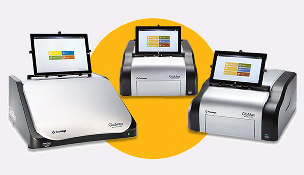
Our cell health assays are compatible with standard plate readers and luminometers. We offer both benchtop options in the GloMax® line or a more compact and portable option with the MyGlo® Instrument.
From Each Well
The GloMax® line includes high-performance plate readers and luminometers with a range of detection options for cell health.
From advanced multimode luminescence, fluorescence, absorbance, BRET and FRET capabilities to dedicated microplate or single-tube luminometers, there is a GloMax® system for every lab and throughput need.
From Start to Finish
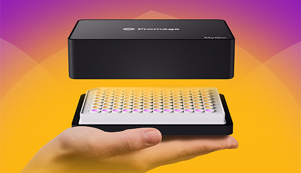
The MyGlo® Instrument is a compact, portable 96-well luminescence plate reader designed for flexibility in modern labs.
The MyGlo® Instrument connects seamlessly to the cloud-based ProNect® Data Platform, which is accessible through any web browser, giving you an end-to-end data solution for your cell health research.
FAQ
What are the main differences between CellTiter-Glo® 2.0 and the original CellTiter-Glo® Assay?
The CellTiter-Glo® 2.0 Assay uses a single, ready-to-use liquid reagent that eliminates the substrate reconstitution step required with the original two-component kit. It also offers improved stability at 4°C over multiple days, making it better suited for repeated use across extended screening campaigns. Assay sensitivity, dynamic range, and signal half-life are equivalent between versions.
| Feature | CellTiter-Glo® (Original) Assay | CellTiter-Glo® 2.0 Assay |
|---|---|---|
| Reagent format | Two-component: lyophilized substrate + buffer (requires reconstitution) | Single, ready-to-use solution (no reconstitution required) |
| Preparation time | ~30min (substrate reconstitution + equilibration) | ~15min (equilibration only) |
| Reagent stability (4°C) | Reconstituted: 48h (~5% loss); 4d (~20% loss) | Stable for repeated use over multiple days at 4°C |
| Sensitivity | Detects as few as 15 cells/well (384-well format) | Detects as few as 15 cells/well (384-well format) |
| Linear dynamic range | Up to 5 logs | Up to 5 logs |
| Signal type | Glow-type; >5h half-life | Glow-type; >5h half-life |
| HTS/automation | Yes: compatible with 96/384/1536-well formats | Yes: optimized for repeated batch use and automation |
| Multiplex compatibility | Standard | Improved; recommended for multiplex workflows |
| Best use case | Standard single-run viability and cytotoxicity assays | High-throughput, multi-day screening campaigns, automation |
Is the sensitivity of the CellTiter-Glo® 2.0 Assay the same as the original?
Yes. The CellTiter-Glo® 2.0 Assay retains the same high sensitivity as the original assay—capable of detecting as few as approximately 15–100 cells/well depending on plate format—with a linear dynamic range spanning up to 5 logs. The reformulation focused on improving stability and workflow, not changing core assay chemistry.
How stable is the CellTiter-Glo® 2.0 Assay compared to the original?
The CellTiter-Glo® 2.0 Assay is stable for repeated use over multiple days when stored at 4°C after opening. The original CellTiter-Glo® Reagent, once reconstituted, is stable for 48 hours at 4°C with approximately 5% signal loss, or 4 days with approximately 20% signal loss—requiring preparation close to the time of use.
Which version is recommended for high-throughput screening?
The CellTiter-Glo® 2.0 Assay is the recommended version for high-throughput screening and automated workflows. The ready-to-use format is more compatible with robotic liquid handlers, reduces batch-to-batch preparation variability, and maintains consistent performance across multi-day plate runs without requiring fresh reagent preparation each session.
Can the CellTiter-Glo® 2.0 Assay be used with 3D cell cultures?
The CellTiter-Glo® 2.0 Assay is not optimized for 3D spheroid or organoid models. For 3D cultures, Promega recommends CellTiter-Glo® 3D Cell Viability Assay, which uses an enhanced lysis buffer specifically formulated to penetrate 3D tumor spheroid structures and release ATP from cells throughout the spheroid.
What is the best cell viability assay for high-throughput drug screening?
ATP-based luminescence assays, such as the CellTiter-Glo® 2.0 Assay, are the preferred platform for HTS drug screening because they offer the highest sensitivity (detecting 15–100 cells/well), the simplest protocol (single reagent addition, 10-minute read time), and the lowest coefficient of variation across plate formats. Colorimetric assays (MTT, MTS) require additional processing steps and offer lower sensitivity at miniaturized volumes.
Are there cost differences between CellTiter-Glo® 2.0 and the original?
List pricing for CellTiter-Glo® 2.0 is modestly higher than the original per volume, reflecting the improved formulation and ready-to-use format. However, for high-throughput laboratories, the time savings from eliminated reconstitution steps and the ability to use reagent across multiple days may offset the per-assay cost difference. Contact your Promega representative for volume pricing options.
What should I do if CellTiter-Glo® 2.0 signals are inconsistent between plates?
Inconsistent signals across plates are most commonly caused by temperature differences between the plate and reagent at the time of addition (ensure both are equilibrated to room temperature for 30 minutes before mixing), insufficient cell lysis (increase mixing time or speed), or signal read outside the stable window (read plates within 10 minutes to 1 hour after stabilization). Confirm that white-walled plates are in use, as plate color significantly impacts luminescence signal intensity.
Resources
AACR 2026 Poster
See screening performance data across suspension, adherent, and 3D cancer cell models
Assays and Tools to Monitor Biology in 3D Cell Cultures
View our practical guide to transitioning cell health assays from 2D to 3D.
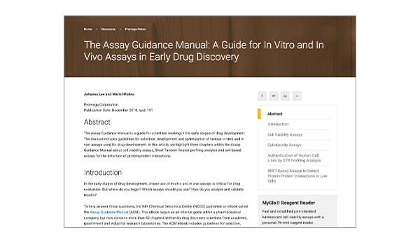
Assay Guidance Manual
Explore step-by-step protocols and recommendations for selecting and running cell health assays.
Interactive Assay Guide
Choose the right cell health assay based on your experimental question, model type, and readout requirements.